
Basal Cell and Squamous Cell Carcinoma: Treatment Options in the UK
Basal cell carcinoma (BCC) and squamous cell carcinoma (SCC) are the two main forms of non-melanoma skin cancer. Both usually develop in areas exposed to ultraviolet radiation, such as the face, ears, scalp, neck, hands and lower legs. BCC is the more common type and generally grows slowly. It very rarely spreads elsewhere in the body, but it can damage nearby skin, cartilage or bone if allowed to enlarge. SCC can also often be cured, although a small proportion behave more aggressively and can spread to nearby lymph nodes or, rarely, more distant organs.
Treatment in the UK is planned according to the cancer’s type, size, depth, position and appearance under the microscope, as well as a person’s general health and preferences. A small superficial BCC on the trunk may be suitable for a cream or light-based treatment, while an SCC on the ear may need surgery and closer follow-up. Most care is delivered through NHS dermatology, plastic surgery or specialist skin cancer services, with complex cases discussed by a multidisciplinary team.
Diagnosis and Factors That Shape Treatment Choices
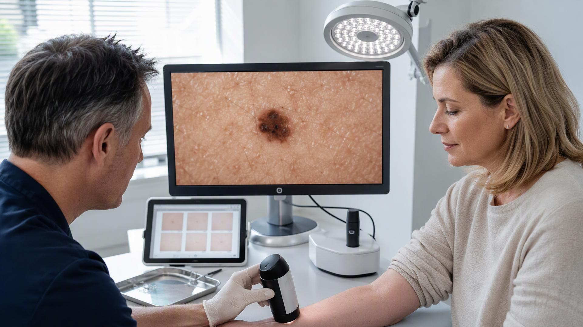
A GP may refer a suspicious lesion through the urgent suspected-cancer pathway, particularly if it is growing, bleeding, crusting, painful or failing to heal. A BCC can appear as a pearly or shiny lump, sometimes with small visible blood vessels, while an SCC may look like a persistent scaly, crusted or tender patch. These appearances can overlap with harmless skin conditions, so examination by a trained clinician is needed.
Diagnosis is commonly confirmed with a biopsy. This means removing all or part of the abnormal area under local anaesthetic and sending it to a laboratory. A pathologist examines the tissue under a microscope to identify the cancer type and features linked with a higher chance of recurrence, such as deeper growth or a less clearly defined edge.
Doctors classify lesions as lower or higher risk rather than offering every patient the same treatment. Location matters because skin cancers on the nose, eyelids, lips, ears and other parts of the central face can be difficult to remove while preserving function and appearance. A tumour that has returned after earlier treatment, has indistinct borders, is large, or occurs in someone with a weakened immune system may also need a more specialised approach.
For people with higher-risk, recurrent or cosmetically sensitive lesions, Dr James Wilson skin cancer treatment includes specialist assessment and non-surgical radiotherapy options where these may be appropriate.
For SCC, assessment includes looking for signs that it has travelled beyond the original skin site. The clinician may examine nearby lymph nodes, such as those in the neck, armpit or groin. Scans are not routinely needed for every small SCC, but may be arranged if there are higher-risk features or symptoms suggesting more extensive disease. BCC rarely needs this type of staging because spread is exceptionally uncommon.
Surgical Removal and Specialist Mohs Surgery
Standard surgical excision is the main treatment for many BCCs and SCCs. The surgeon removes the visible cancer along with a small rim of normal-looking skin, known as a margin. This provides a better chance of clearing microscopic cancer cells at the edge of the lesion. The specimen is then examined by a laboratory to check whether the margins are clear.
Most procedures are carried out using local anaesthetic, meaning the patient stays awake but the area is numbed. Smaller wounds may be closed with stitches, while larger ones may heal naturally, be covered with a skin graft, or require a flap of nearby skin to be moved into place. The choice depends on the site, wound size and how much tension there would be on the surrounding skin.
Curettage and cautery can be used for selected low-risk lesions. The doctor scrapes away the cancer with a sharp instrument, then applies heat to stop bleeding and destroy remaining surface cells. It is less suitable for cancers with uncertain boundaries or those in cosmetically sensitive, high-risk areas, because it does not provide the same detailed margin assessment as excision.
Mohs micrographic surgery is a precise technique used mainly for complex BCCs and selected SCCs. The surgeon removes thin layers of tissue one at a time and examines each layer under a microscope during the appointment. More tissue is taken only where cancer cells remain. This can preserve as much healthy skin as possible, which is particularly valuable around the eyes, nose, lips, ears and fingers, or when a cancer has recurred.
Non-Surgical Treatments for Selected Skin Cancers
Radiotherapy uses carefully targeted high-energy X-rays to destroy cancer cells. It may be offered when surgery would be difficult because of the lesion’s location, when a person is not well enough for an operation, or after surgery if an SCC has features that raise the risk of return. Treatment is usually given in several sessions. The skin may become red, sore or flaky during treatment, and longer-term changes can include altered skin texture or colour.
Photodynamic therapy, often called PDT, is mainly used for superficial BCC and certain precancerous sun-damaged areas. A light-sensitive cream is applied to the lesion and allowed time to be absorbed. A special light then activates the cream, damaging the abnormal cells. PDT can be helpful where several superficial areas need treatment, although the treated area may be painful, red and crusted for a period afterwards.
Topical medicines are another option for certain surface-level BCCs. Imiquimod stimulates a local immune response, while 5-fluorouracil interferes with the growth of abnormal cells. These creams are prescribed for a set course and often cause temporary inflammation, redness, soreness and scaling, which can indicate that the treatment is working. They are not appropriate for deeper, infiltrative or high-risk cancers, so a confirmed diagnosis and specialist advice are necessary.
Advanced Disease, Follow-Up and Skin Protection
Advanced BCC and SCC are uncommon, but specialist treatment is available when cancer cannot be fully removed or has spread. Management may combine surgery and radiotherapy. For advanced SCC, immunotherapy drugs can help the immune system recognise and attack cancer cells in suitable patients. Advanced BCC may be treated with targeted medicines that block signals involved in BCC growth. These treatments require assessment by an experienced cancer team because side effects and expected benefits vary.
Follow-up arrangements are tailored to risk. People treated for a straightforward low-risk BCC may be discharged after treatment with advice on checking their skin. Higher-risk BCC, advanced disease, multiple tumours and immune suppression may lead to planned review appointments. Following SCC, specialist follow-up is more likely when the tumour had high-risk features, as clinicians need to check the original site and nearby lymph nodes for signs of recurrence.
New skin cancers are possible after a previous BCC or SCC, so regular self-checking is useful. Seek medical advice about a new or changing mark, a sore that does not heal, a scab that repeatedly returns, or any change around a treated area. Reducing ultraviolet exposure also lowers future risk: use shade, protective clothing and a broad-brimmed hat, avoid sunbeds, and apply high-protection sunscreen to exposed skin when appropriate.
A Practical Route to Effective Care
BCC and SCC are often highly treatable, especially when assessed early and managed with a treatment matched to their individual features. Surgery remains the most common option, but radiotherapy, creams, PDT and specialist medicines all have defined roles. Promptly reporting a persistent or changing skin lesion, attending follow-up when advised and protecting skin from further ultraviolet damage give patients the strongest basis for successful treatment and long-term skin health.